Nub theory is the most popular way to guess a baby’s sex from a 12-week scan photo, and this whole piece is for fun. It’s a parlour game with a kernel of actual anatomy behind it. Nothing here is medical advice or a reading of your own scan; the people who can tell you the sex with any confidence are the sonographer at your anatomy scan or the clinician reading a blood test.
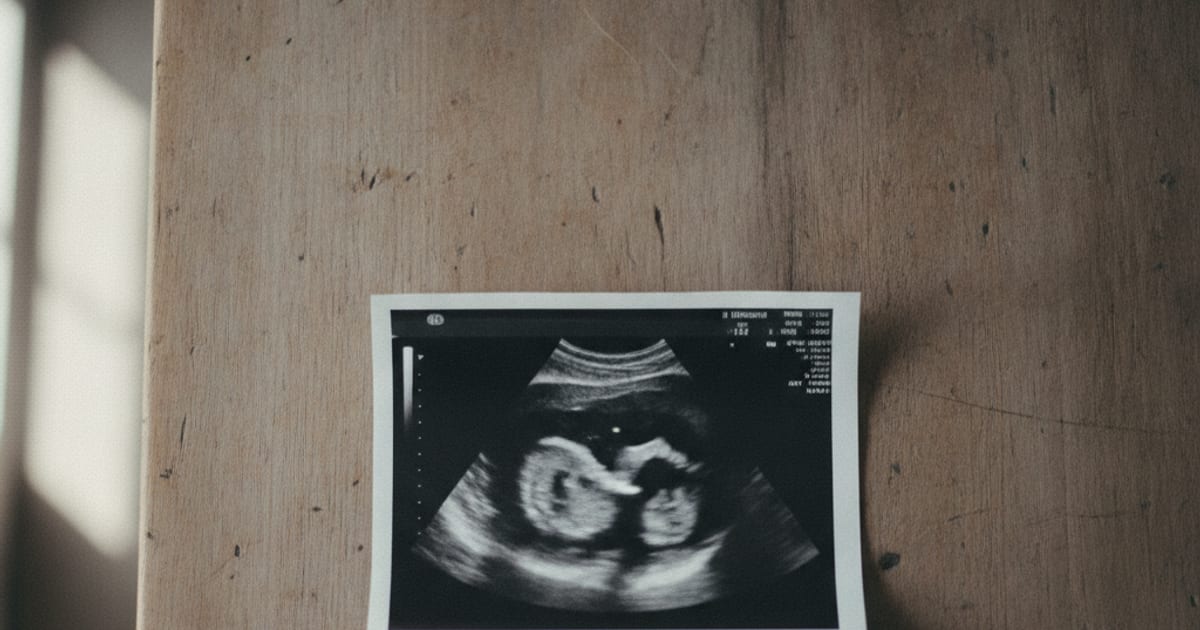
I first heard the term in a group chat the night after my own 12-week scan, when someone asked me to send the printout so they could “check the nub”. Here’s what they were looking at, how people read it, and what the research says (more interesting than the group chat admitted).
Nub theory says you can guess a baby’s sex at the 11 to 14 week scan from the angle of the genital tubercle, a small bump that later becomes the penis or clitoris. Roughly parallel to the baby’s back suggests a girl; tilted up 30 degrees or more suggests a boy. It’s a for-fun method, never a diagnosis.
What nub theory is (and what the nub actually is)
Between about 11 and 14 weeks, every baby has the same small bump between the legs. Embryologists call it the genital tubercle. It develops into a penis in boys and a clitoris in girls, but in that window the two look nearly identical in size and shape.
What differs, on average, is the direction it points. The sonographers who studied this in the late 1990s drew a line along the skin of the baby’s lower back (the lumbosacral skin surface, in the papers) and measured the angle between that line and the tubercle in a side-on view.
In the original 1999 protocol, an angle greater than 30 degrees was called male, and a tubercle lying parallel to the line or dipping toward it was called female. Later protocols (2006 and 2012) tightened that: over 30 degrees male, under 10 degrees female, and 10 to 30 degrees “not determined”. Keep that middle zone in mind, because the researchers wouldn’t call it and the internet always does.
The community vocabulary is its own thing. A “stacked” nub means a boy nub with a bright line layered on top of it, and “forked” means the tip looks split in two (readers argue about what that means). Neither term comes from the research; they’re folk descriptions of a 2D image, and part of the fun.
How to read nub theory on a 12 week scan photo
You need a clear profile: the baby lying side-on with the spine running flat across the picture (the midsagittal plane, in sonographer terms), the same view used for the crown-to-rump measurement. If the baby is facing you, lying at an angle or curled into a C-shape, any angle you measure is meaningless.
Then find the nub, a small bump at the bottom of the torso where the legs begin, a little in front of the base of the spine. Lying roughly level with the lower back, or pointing slightly down toward the feet, is the girl pattern. Tilting up toward the head at something like 30 degrees or more is the boy pattern, and anything in between is a genuine no-call, whatever the group chat says.
Plenty of photos can’t be read at all. The usual culprits:
- A face-on or three-quarters view, so there’s no true profile.
- Legs crossed or pulled up over the nub.
- The umbilical cord running between the legs and passing for a nub.
- A 3D or 4D render instead of a 2D grey-scale frame.
- A scan taken before roughly 12 weeks, when many boy nubs haven’t tilted yet.
- A zoomed-out, grainy frame with the lower body in shadow.
Even in the published studies, where a sonographer could move the probe and wait for the baby to stretch out, the view wasn’t always there. The 2006 Israeli team assigned a sex in 613 of 656 fetuses (93%). Your single frozen frame has worse odds of being usable than their live scan did.
Nub theory accuracy: what the research actually found
In 1999, Efrat, Akinfenwa and Nicolaides at King’s College Hospital scanned 172 singleton pregnancies at 11 to 14 weeks and called the sex from the genital tubercle angle in a midsagittal view, using 30 degrees as the boy threshold. Accuracy rose with gestation: 70.3% at 11 weeks, 98.7% at 12 weeks and 100% at 13 weeks. The error ran one way: at 11 weeks, 56% of boys were wrongly called girls, while only 5% of girls were called boys.
In 2006, Efrat and colleagues at Rabin Medical Center repeated the exercise in 656 pregnancies at 12 to 14 weeks, leaving angles of 10 to 30 degrees undetermined. A sex was assignable in 93% of fetuses. Checked against 555 newborns, boy calls were 99 to 100% correct at every age, while girl calls were 91.5% correct at 12+0 to 12+3 weeks, 99% at 12+4 to 12+6, and 100% from 13 weeks.
Two things jump out. Accuracy climbs steeply with every few days of gestation, and before 12 weeks the method is weak, mostly because boy nubs haven’t tilted yet.
Later work was less rosy. A 2012 University of Colorado study of 260 fetuses at 11 to 13+6 weeks, using the same cut-offs, assigned a sex in 93.1% and got it right in 85.8% (boys 91.4%, girls 80.5%). Higher maternal BMI made the image harder to get, though it didn’t lower accuracy once a usable image existed.
A 2025 systematic review in the Journal of Ultrasound pooled 28 studies. Across the 9 first-trimester studies the mean accuracy was 85%, and pooled sensitivity climbed from 69% at 11 weeks to 89% at 12 and 96% at 13. Second- and third-trimester scans ran at a median of 99%.
Why a printout read is different from a sonographer’s measurement
Every figure above came from a trained operator holding the probe, choosing the plane, waiting for a straight profile and measuring the angle on the machine. That’s the method the studies tested.
A nub read on a phone photo of a printout is a different exercise. The frame was frozen for the crown-rump measurement, with no thought for the genitals, and the angle gets eyeballed from a picture that may sit slightly off-plane, which changes the apparent tilt. And the 10-to-30-degree zone the 2006 and 2012 protocols refused to call is exactly where most “it’s leaning, so boy” arguments live.
There’s also the coin. Guessing gets you about 50% for free, so any method (a Facebook group, a chatbot, your aunt) will “work” half the time. For a reading to mean something it would have to beat that across many babies, and I couldn’t find a published study that tested community nub readings at all.
What about the ChatGPT nub theory?
People now paste their printout into a chatbot and ask. Treat the answer like a confident friend’s: it’s reading one frozen 2D frame with no measurement, no crown-rump length and no second view. I couldn’t find a peer-reviewed study on chatbot nub readings, and the sonographer figures above don’t transfer to a screenshot.
Nub theory vs Ramzi theory vs the Chinese gender chart
The popular guessing games stand on very different ground.
- Nub theory (11 to 14 weeks) has an anatomical basis and published accuracy figures, but only for sonographer measurements at the right gestation.
- Ramzi theory (6 to 8 weeks) claims the side the placenta forms on predicts the sex. When I searched PubMed for the original claim, nothing came up; as far as I can tell it was never published in a peer-reviewed journal.
- The Chinese gender chart matches the mother’s lunar age to the month of conception. Villamor and colleagues tested it against 2,840,755 Swedish births from 1973 to 2006 and found agreement no better than a coin toss (kappa 0.0002). You can still play with our Chinese gender chart; it’s there for fun too.
The reliable ways to learn the sex
When you want an answer you can paint a room on, these are the routes.
The anatomy scan. In the UK the NHS offers the mid-pregnancy scan between 18 and 21 weeks, and you may be asked whether you’d like to know the sex. It’s a screening scan of how the baby is developing; the sex is a bonus, and the sonographer may not get a clear view on the day. US practices run a second-trimester anatomy scan on a similar schedule, so ask your provider when yours falls.
Cell-free DNA screening (cfDNA, often called NIPT). A blood draw from the mother picks up fragments of the baby’s DNA, and finding Y-chromosome sequences points to a boy. A 2011 meta-analysis in JAMA of 57 studies (6,541 pregnancies) found 95.4% sensitivity and 98.6% specificity for detecting those sequences, with tests before 7 weeks described as unreliable and performance best after 20 weeks. It’s ordered through a clinician, and some direct-to-consumer kits use the same principle; your provider can tell you what’s appropriate for you.
Whatever route you take, the person to ask about your own pregnancy is your midwife or doctor.
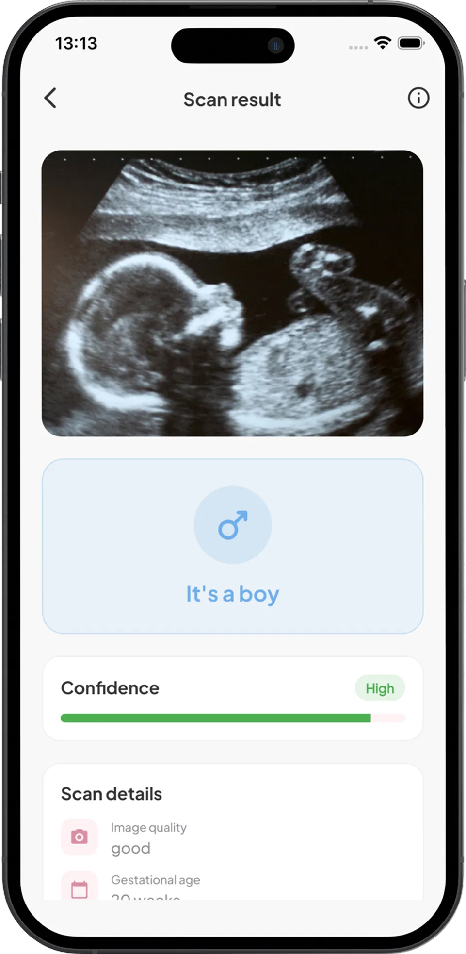
How Baby Gender Scanner: Nubbi helps (for fun, after 14 weeks)
Nub theory closes its window at around 14 weeks, which is exactly where Baby Gender Scanner: Nubbi opens its own. Upload or snap any ultrasound photo taken at 14 weeks or later, and in a few seconds it gives a boy-or-girl prediction with a confidence score. Think of it as the group-chat guess with a number pinned to it.
A few honest notes. It’s built for fun and curiosity, and the result is a pattern-matching guess; it doesn’t work on photos taken before 14 weeks, so your 12-week nub shot stays with the group chat. For a clinical answer you still need a doctor, midwife or sonographer.
Privacy is simple: images are processed on the fly and never kept on servers. The free version gives you the guess. Premium adds the full write-up, “what the AI spotted” markers on your image, unlimited predictions and a pregnancy timeline that lines up your scans as the weeks go by.
I use it the way I used nub theory in that group chat, as a reason to stare at the picture a little longer and argue about it.
Nub theory FAQ
How can you tell if a nub is a boy or girl?
In a true side profile, compare the direction of the small bump between the legs with the line of the lower back. Lying level with it, or dipping down, is the girl pattern; tilting up at about 30 degrees or more is the boy pattern. Between 10 and 30 degrees the research protocols made no call, and neither should you.
What does a 12 week boy nub look like?
In the published method, a boy nub points up toward the head at more than 30 degrees from the line of the lower back. On printouts, readers also look for a bright second line layered over the nub, which they call stacking. At 11 weeks many boy nubs haven’t tilted yet; in the 1999 study 56% of boys were wrongly called girls at that stage.
How accurate is the ChatGPT nub theory?
I couldn’t find a published study testing chatbot nub readings, so there’s no accuracy figure to quote. A chatbot sees one frozen 2D frame with no angle measurement and no check on gestational age. A coin gets 50% for free; treat the chatbot’s answer as the same kind of fun.
When is nub theory most accurate?
The later in the window, the better. In Efrat’s 1999 series accuracy was 70.3% at 11 weeks, 98.7% at 12 and 100% at 13, and the 2025 meta-analysis of 28 studies found sensitivity rising from 69% at 11 weeks to 96% at 13. Those figures are for live sonographer measurements, not photo reads.
Is nub theory less accurate for girls?
In the studies, girl calls were the weaker ones early on. The 2006 series found 91.5% accuracy for girl calls at 12+0 to 12+3 weeks versus 99 to 100% for boys, and the 2012 study found 80.5% for girls versus 91.4% for boys. The usual reason is a boy nub that hasn’t tilted yet and looks like a girl nub.
If your 12-week printout turned into a family debate, the next scan will be even more fun. Once you have a photo from 14 weeks or later, run it through Baby Gender Scanner: Nubbi for a boy-or-girl guess with a confidence score, then compare notes at the anatomy scan. It’s a bit of play for the early pregnancy stage; the real reveal still belongs to your sonographer or clinician.
Sources
- Efrat Z, Akinfenwa OO, Nicolaides KH. First-trimester determination of fetal gender by ultrasound. Ultrasound Obstet Gynecol 1999;13(5):305-307. PubMed 10380292
- Efrat Z, Perri T, Ramati E, Tugendreich D, Meizner I. Fetal gender assignment by first-trimester ultrasound. Ultrasound Obstet Gynecol 2006;27(6):619-621. PubMed 16493625
- Behrendt N, Foy P, Center J, Durnwald CP. Influence of maternal body mass index and gestational age on accuracy of first trimester gender assignment. J Matern Fetal Neonatal Med 2012;25(3):253-256. PubMed 21500978
- Amankona E, Donkor A, Agyei BA, Anyitey-Kokor I, Odoi AT, Wiafe YA. Effectiveness of prenatal ultrasound in fetal sex identification: a systematic review and meta-analysis. J Ultrasound 2025;28(3):563-574. PubMed 39798051
- Devaney SA, Palomaki GE, Scott JA, Bianchi DW. Noninvasive fetal sex determination using cell-free fetal DNA: a systematic review and meta-analysis. JAMA 2011;306(6):627-636. PubMed 21828326
- Villamor E, Dekker L, Svensson T, Cnattingius S. Accuracy of the Chinese lunar calendar method to predict a baby’s sex: a population-based study. Paediatr Perinat Epidemiol 2010;24(4):398-400. PubMed 20618730
- NHS. 20-week screening scan. Accessed 2026-10-04.